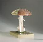
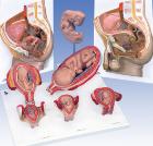
Model For Gynecological Patient Education, This unique gynecological training model is ideal for demonstration purposes and for realistic insertion of female barrier contraceptive devices, which are placed in the vaginal/cervical area.
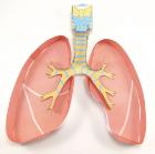
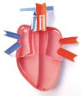
Human Anatomy Transparent Torso Model 4D, 8in model contains 37 detachable, hand-painted medical education-quality organs and parts and a display stand, Also includes illustrated assembly guide and description of the anatomy, For ages 8 plus
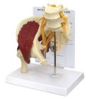
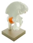
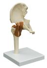
Model of human hip joint. With simulated pubofemoral ligament, Mounted on base, represents the upper femur and pelvic bone and the way that they move, Numbered with English Key Card, Size 22 x 12 x 25 cm approx. Weight 800 g approx
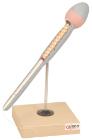
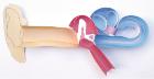
Simulator Catheterisation-Model, male, Ideal for demonstrating disposable & ballon catheters as well as suprapubic aspiration. Can be dismantled into two parts, natural size, made of special plastic. On a stand with green base, LxWXH: 18 X 18 X 30 cm
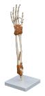
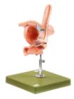
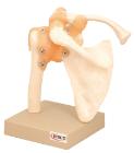
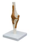
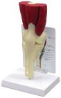
Model of human elbow joint. Mounted on base. Features upper half of radius and ulna, lower half of humerus and the ligaments of the elbow joint, to show hinge joint, Numbered with English Key Card, Size 17x 22 x 22 cm approx, Weight 250 g
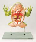
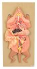
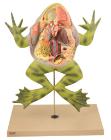
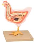
Model showing life like sculpting and vibrant hand painting faithfully portray ten organ systemsr, with numbered features. Molded of non breakable plastic base, bull frog lifts off its plastic stand for hands on study
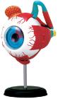
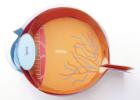
Human Anatomy Eyeball Model 4D, 4.5in model contains 35 detachable, hand-painted medical education-quality organs and parts and a display stand, Also includes illustrated assembly guide and description of the anatomy, Offer a unique puzzle challenge, For ages 8 plus
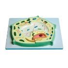
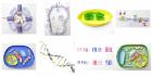
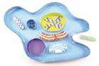
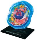
Science Animal Cell Model 4D, 6in model contains 26 detachable, hand-painted parts and a display stand, Transparent outside for seeing internal structures and super detailed parts, Also includes illustrated assembly guide, description of the anatomy, For Ages 8 plus
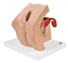
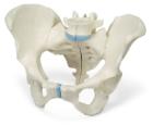
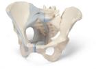
Female Pelvis 3 Part, This life size three part model represents an original cast of a bony female pelvis and shows all the details of the following anatomical structures